 Breast Surgical Oncologist
Advanced Surgical Services - By Dr. Neemesh Lodh
- Services
- Breast Cancer Surgery (Early & Advanced Stage)
- Breast Conserving Surgery (Lumpectomy)
- Oncoplastic Breast Surgery & Reconstruction
- Skin-Sparing & Nipple-Sparing Mastectomy
- Sentinel Lymph Node Biopsy & Axillary Surgery
- Mammary Ductoscopy for Nipple Discharge
- Breast Lump Evaluation & Biopsy
- Management of Benign Breast Diseases
- Gynecomastia (Male Breast) Surgery
- Chemoport Insertion for Chemotherapy
- Genetic Counseling & Risk Reduction Surgery
- High-Risk Breast Cancer Screening Programs
- Multidisciplinary Breast Cancer Care Planning
Breast Lump Evaluation & Biopsy
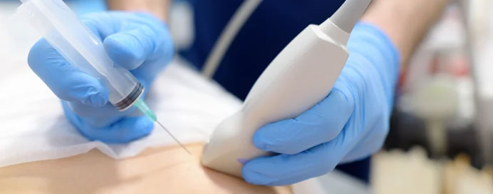
Breast lump evaluation is a systematic process used to determine the nature of a breast lump—whether it is benign (non-cancerous) or malignant (cancerous). Early and accurate assessment is essential for proper diagnosis and timely treatment. Evaluation typically includes clinical examination, imaging studies such as ultrasound or mammography, and, when required, a biopsy to obtain tissue for laboratory analysis. A biopsy is the most definitive method to diagnose the exact cause of a breast lump and helps guide further management, ensuring personalized and effective care.
Key Components of Breast Lump Evaluation & Biopsy
- Detailed Clinical Examination by a Specialist: A thorough physical examination is the first step in evaluating a breast lump. The doctor assesses the size, shape, consistency, mobility, tenderness, and location of the lump, along with checking for skin changes, nipple discharge, or lymph node enlargement. A detailed medical history—including age, family history of breast cancer, hormonal history, and duration of the lump—helps determine the level of suspicion and the next appropriate diagnostic steps.
- Advanced Imaging for Accurate Assessment: Imaging studies play a crucial role in differentiating between solid and cystic lumps. Ultrasound is commonly used in younger women or for further evaluation of a palpable lump, while mammography is preferred in women above 40 years of age. In selected cases, breast MRI may be recommended for detailed visualization. Imaging helps categorize the lump using standardized reporting systems and guides whether a biopsy is necessary.
- Minimally Invasive Biopsy Techniques for Definitive Diagnosis: If imaging or clinical findings raise suspicion, a biopsy is performed to obtain a tissue sample. Common methods include Fine Needle Aspiration Cytology (FNAC), Core Needle Biopsy, and Vacuum-Assisted Biopsy. Core needle biopsy is most commonly used as it provides sufficient tissue for accurate histopathological examination. These procedures are typically done under local anesthesia and are safe, quick, and minimally painful.
- Histopathological Analysis for Precise Results: The collected tissue sample is examined under a microscope by a pathologist to determine whether the lump is benign, pre-cancerous, or malignant. In cases of cancer, additional tests such as hormone receptor status (ER, PR) and HER2 testing may be performed. These results are essential in planning the most appropriate treatment, including surgery, chemotherapy, hormonal therapy, or targeted therapy.
- Personalized Treatment Planning and Follow-Up Care: Based on the biopsy findings, a customized treatment plan is created. Benign lumps may only require observation or minor surgery, while malignant lumps require comprehensive cancer management. Regular follow-up and monitoring are essential to ensure successful outcomes and early detection of any recurrence. Early evaluation and biopsy significantly improve prognosis and treatment success rates.